

De hersenen
Algemeen
Overzicht
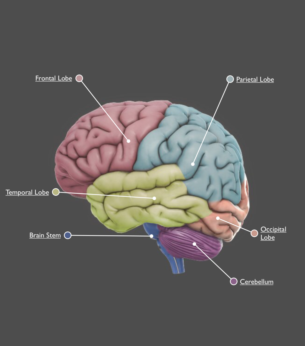
De hersenen zijn een unieke structuur die bepaalt wie we zijn als individuen en hoe we de wereld ervaren. Door de recente vooruitgang op het gebied van “neuroimaging” konden onderzoekers in de hersenen kijken en levendige beelden van de subcomponenten bekijken en de bijbehorende functies benoemen. De grove structuur van de hersenen is bij de meesten bekend. De buitenste laag van de voorhersenen vormt het bekende gerimpelde weefsel dat de hersenschors of kortweg de cortex is. De grote plooien in de cortex worden gyri genoemd (uit het Grieks ‘cirkel’). De kleine vouwen in deze plooien zijn sulci (uit het Grieks, ‘geul’). Elke hersenhelft van de cortex bestaat uit vier lobben – frontaal, pariëtaal, temporaal en occipitaal. Andere belangrijke structuren zijn de hersenstam, het cerebellum het limbisch systeem (waaronder de amygdala en de hippocampus).
Hieronder bespreken we 28 structuren. Elke structuur bevat informatie over bijbehorende functies, stoornissen, hersenschade, casestudy’s en links naar hedendaags modern onderzoek.
Case studies
Phineas Gage is misschien wel de meest bekende casus in de neurowetenschappen. Hij liep na een spoorweggerelateerd ongeval in 1848 ernstige schade aan de prefrontale cortex op. Een explosie dreef een grote ijzeren staaf door de schedel van Gage en later werd gemeld dat hij ernstige sociale beperkingen vertoonde. Hoewel de omvang van deze stoornissen controversieel blijft, was zijn casestudy een mijlpaal omdat het specifieke cognitieve functies correleerde met een specifiek hersengebied. Sindsdien hebben tienduizenden casestudy’s geprobeerd om specifieke hersenregio’s te associëren met specifieke functies, en veel daarvan hebben we hier gedocumenteerd. Het is echter belangrijk om hier niet in te overdrijven.
Alle cognitieve functies zijn het resultaat van de integratie van vele eenvoudige verwerkingsmechanismen, verspreid over de hersenen.
Bijbehorende functies
- opwinding
- emotie
- taal
- leren
- geheugen
- beweging
- perceptie
- sensatie
- denken
- vele anderen
Bijbehorende cognitieve stoornissen
Almost without exception, cognitive disorders correlate to multiple regions in the brain. Just as the genes and biochemicals associated with cognition are expressed throughout the brain, gross structures that correlate with cognitive disorders are widespread. This is certainly true of the six disorders covered in G2C Online: ADHD, Alzheimer’s disease, autism, bipolar disorder, depression, and schizophrenia.
Bijna zonder uitzondering correleren cognitieve stoornissen met meerdere hersengebieden. Net zoals de met cognitie geassocieerde genen en biochemicaliën door de hersenen worden uitgedrukt, zijn grove structuren die correleren met cognitieve stoornissen wijdverbreid. Dit geldt zeker voor de zes aandoeningen die hier worden behandeld: ADHD, de ziekte van Alzheimer, autisme, bipolaire stoornis, depressie en schizofrenie.
Geassocieerd met schade
De hersenen kunnen beschadigde neurale netwerken herstellen of het functieverlies in bepaalde structuren compenseren. Veelvoorkomende stoornissen als gevolg van hersenbeschadiging zijn onder meer tekortkomingen in aandacht, emotie, taal, leren, geheugen, beweging, perceptie en gevoel.
Substructuren
- amygdala
- basale ganglia
- hersenstam
- Broca’s gebied
- cerebellum
- cingulate cortex
- corpus callosum
- dentate gyrus
- entorinale cortex
- frontale kwab
- hippocampus
- hypothalamus
- inferieure temporale gyrus
- limbisch systeem
- medulla
- middelste temporale gyrus
- occipitale / achterhoofskwab
- pariëtale kwab
- perirhinal cortex
- pons
- prefrontale cortex
- premotorische cortex
- primaire motorische cortex
- somatosensorische cortex
- subiculum
- superieure temporale gyrus
- temporale kwab
- thalamus
- ventrikels
- gebied van Wernicke
- en vele anderen
Onderzoeksrecensies
- Gae (2013) examine the affect of a schizophrenia susceptibility gene on cognitive deficits of neural developmental disorder and schizophrenia (PMID: 23543703).
- Oe and colleagues (2013) review the effects of long-term potentiation on spine density in rodents, modeling the system of memory development (PMID: 23739837). Synaptogenesis spikes during stimulation in a stochastic manner, and is inversely related to the synaptic density of the dendrites.
- Wishart and colleagues (2012) identify six regulators that alter neurodegeneration after injury in Drosophila and mice (PMID: 22952455).
- Casillas–Espinosa and colleagues (2012) review the different mechanisms involved in synaptic transmission and their role in epilepsy (PMID: 23216578).
Links
Amygdala
Overzicht
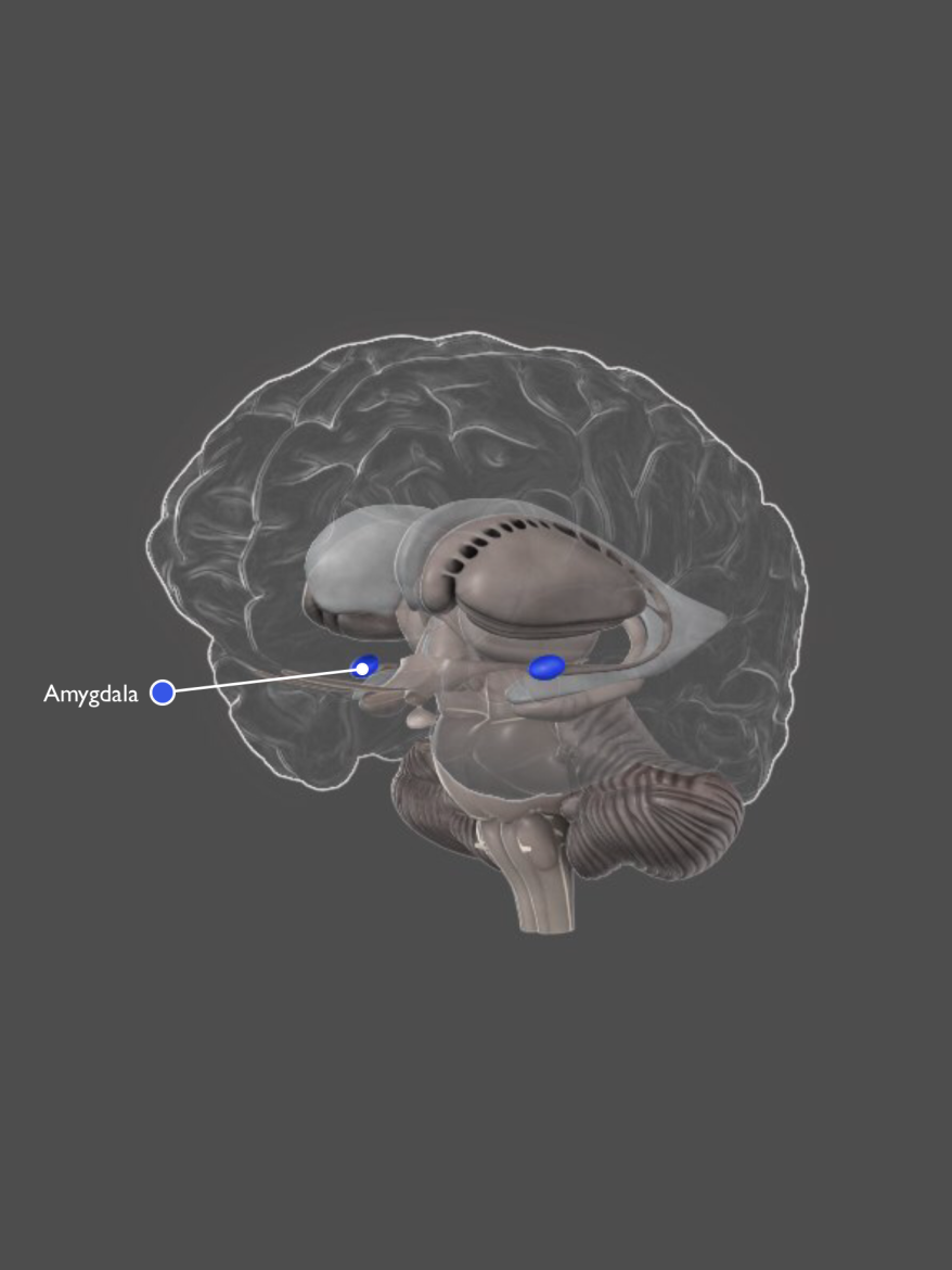
De amygdala is een complexe structuur naast de hippocampus. De amygdala is betrokken bij het verwerken van emoties en het leren van angst. Het verbindt gebieden van de cortex die ‘hogere’ cognitieve informatie verwerken met hypothalamus- en hersenstelsels die ‘lagere’ metabole reacties regelen (bijv. aanraking, pijngevoeligheid en ademhaling). Hierdoor kan de amygdala fysiologische reacties coördineren op basis van cognitieve informatie – het bekendste voorbeeld is de vecht- of vluchtreactie.
De amygdala heeft drie functioneel verschillende delen:
1) de mediale groep subnuclei heeft veel verbindingen met de reukbol en de reukcortex,
2) de basolaterale groep heeft uitgebreide verbindingen met de hersenschors, met name de orbitale en mediale prefrontale cortex, en
3) de centrale en voorste groep van kernen heeft veel verbindingen met de hersenstamhypothalamus en sensorische structuren.
Case studie
De neurologische patiënt SM heeft uitgebreide schade aan de amygdala in elk hemisfeer. Ze heeft geen motorische, sensorische of cognitieve stoornissen. Toen haar werd gevraagd om foto’s van een reeks gezichtsuitdrukkingen te identificeren, kon SM elke uitdrukking identificeren, behalve één, ze herkende angst niet. Wanneer SM werd gevraagd gezichtsuitdrukkingen te tekenen, kon ze een tekening maken van elke emotie, maar ze kon de uitdrukking van angst niet reproduceren. Toen haar werd gevraagd naar haar tekeningen, legde ze uit dat ‘ze niet wist hoe een bang gezicht eruit zou zien’.
Bijbehorende functies
- angstverwerking
- emotionele verwerking
- leren
- vecht of vlucht reactie
- beloningverwerking
Associated cognitive disorders
Many studies have linked autism with amygdala dysfunction. The lack of empathy often shown by autistic individuals has associated with the amygdala (Blair, 2008). Neural activity in the amygdala has also been strongly linked to depression (Northoff, 2007) and bipolar disorder (Phillips & Vieta, 2007). There is very strong evidence linking post–traumatic stress disorder with amygdala responses (Brewin, 2008).
Associated with damage
- aggression
- irritability
- loss of control of emotion
- disruption of short–term memory
- deficits in recognizing emotions (particularly fear)
Research reviews
- Blair (2008) discusses two disorders, psychopathy and autism, in relation to abnormal amygdala functioning (PMID: 18038346).
- Brewin (2008) reviews neurological correlates of post–traumatic stress disorder, which has been strongly linked to the amygdala (PMID: 18037017).
- Phillips and Vieta (2007) consider functional abnormalities of the amygdala as potential biomarkers of bipolar disorder (PMID: 17562698).
- Morrison and Salzman (2010) review the different roles of the amygdala, including coordinating affective/emotional responses (PMID: 20299204).
- Northoff (2007) reviews the neuropathology of depression, which has been linked with abnormal neural activity in a number of structures, including the amygdala (PMID: 17379318).
- Roozendaal and colleagues (2009) review studies that found neural correlates of stress–induced modulation of the amygdala (PMID: 19469026).
- Crocker and colleagues (2013) review evidence for cognitive biases and deficits in depression and anxiety, and how they interact with emotional processes (PMID: 23781184).
Links
De basale ganglia
![]()
Overzicht
The basal ganglia comprise a group of structures that regulate the initiation of movements, balance, eye movements, and posture. They are strongly connected to other motor areas in the brain and link the thalamus with the motor cortex. The basal ganglia are also involved in cognitive and emotional behaviors and play an important role in reward and reinforcement, addictive behaviors and habit formation.
Case study
A 2008 study by Das and colleagues that approximately 5% of apparently healthy middle–aged adults have micro–lesions in the basal ganglia. Known clinically as silent cerebral infarcts (SCI), these lesions have an overall prevalence of about 10% in seemingly healthy adults.
Associated functions
- movement regulation
- skill learning
- habit formation
- reward systems
Associated cognitive disorders
The basal ganglia are particularly associated with movement disorders such as Parkinson’s and Huntington’s disease. In terms of cognitive disorders, basal ganglia abnormalities have been found in individuals with schizophrenia, and may explain habit learning deficits associated with the disorder (Keri, 2008). The basal ganglia may also contribute the neuropathology of depression, particularly in relation to the limbic system (Stathis and colleagues, 2007).
Associated with damage
- tremors
- involuntary muscle movements
- abnormal increase in muscle tone
- difficulty initiating movement
- abnormal posture
Substructures
- caudate nucleus
- globus pallidus
- nucleus accumbens
- putamen
- substantia nigra
- subthalamic nucleus
Research reviews
- Das and colleagues (2008) examine neuroimaging data from over 2040 middle–aged participants and find evidence of silent cerebral infarcts in 10.7% of the sample (PMID: 18583555).
- Weber (2012) examines risk of recurrent stroke in adults with silent cerebral infarctions (PMID: 22267825).
- Keri (2008) includes the basal ganglia in a review on category–learning deficits in schizophrenia (PMID: 17854895).
- Foerder and Shohamy (2011) review the role of the basal ganglia in non–declarative memory, motivation, and decision making (PMID: 21945835).
- Stathis and colleagues (2007) review associations between the basal ganglia, limbic system, and cognitive disorders with an emphasis on neuromodulation (PMID: 17691350).
- Yoon and colleagues (2013) suggest a relationship between the prefrontal basal ganglia and schizophrenia (PMID: 23290498).
De herstenstam
Overview
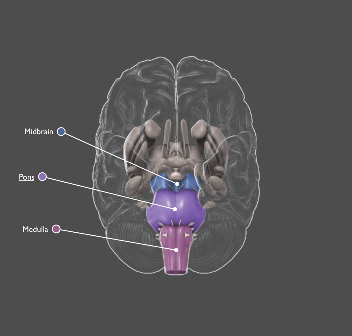
The brain stem consists of a group of structures that lie deep within the brain, including the pons, medulla oblongata, and midbrain. It plays an important role in maintaining homeostasis by controlling autonomic functions such as breathing, heart rate, and blood pressure. While the brain stem can organize motor movements such as reflexes, it coordinates with the motor cortex and associated areas to contribute to fine movements of limbs and the face.
Case study
Cattaneo and colleagues (2006) describe two patients with damage (stroke) to the brain stem who had considerable difficulty balancing and walking. In addition, both were afflicted with “pathological yawning”, which occurred at a frequency of every 2 to 3 minutes.
Associated functions
- maintaining homeostasis by controlling autonomic functions (including blood pressure, breathing, digestion, heart rate, perspiration and temperature)
- alertness
- sleep
- balance
- startle response
Associated cognitive disorders
Very few cognitive disorders have been associated with the brain stem. A number of studies have found brain stem abnormalities in individuals with autism (e.g. Rodier, 2003).
Associated with damage
- organ failure sleep disorders (e.g. insomnia, sleep apnea)
- difficulties balancing and moving
Substructures
- medulla oblongata
- midbrain
- pons
Research reviews
- Cattaneo and colleagues (2006) report two cases of pathological yawning following stroke damage to the brain stem (PMID: 16174652).
- Rodier (2003) reviews evidence for brain stem injury in autism (PMID: 12349873).
- Jou and colleagues (2008) find a significant difference in the gray matter of the brainstem of autistic boys compared with non–autistic boys (PMID: 18812009).
Links
Gebied van Broca
Overview
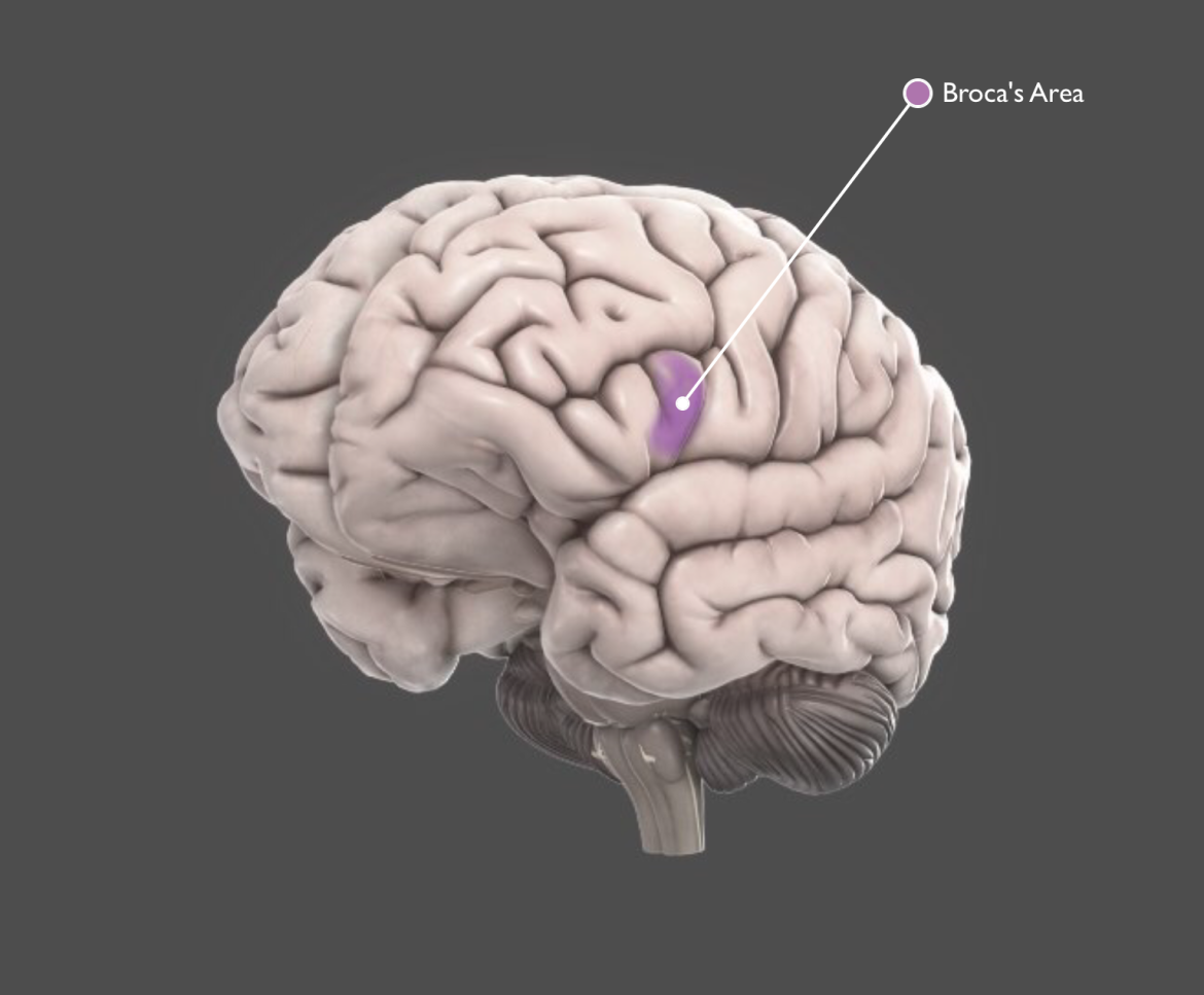
Broca’s area is a functionally defined structure in the left frontal lobe of about 97% of humans (including a large majority of left–handers). Broca’s area is involved mainly in the production of spoken and written language and also in language processing and comprehension. It takes its name from the French scientist whose work with language–impaired patients led him to conclude that we speak with our left–brain.
Case study
In the 1960s, Roger Sperry and Michael Gazzaniga devised an ingenious experiment to determine whether language was lateralized to the left hemisphere. They worked with split–brain patients (patients who had the connective tissue between right and left hemispheres removed). Using the left hemisphere, split–brain patients could produce the name of objects held in the right hand without difficulty. Amazingly, if they held the same object in the left hand (which is controlled by the right hemisphere), the object could not be named.
Associated functions
- language production (both speech and sign)
- comprehension of complex syntax
Associated cognitive disorders
Language impairments in autism may be related to abnormalities in Broca’s area (see Bauman and Kemper, 2005).
Associated with damage
- Broca’s aphasia (inability to express language) which includes halting speech
- repetitive speech (perseveration)
- disordered syntax and grammar and disordered structure of individual words
Research reviews
- Bauman and Kemper (2005) review neuroanatomical evidence that language impairments in autism may be related to abnormalities in Broca’s area (PMID: 15749244).
- Anderson and colleagues (2009) review evidence of lower activity in Broca’s area and Wernicke’s area in high functioning individuals on the autism spectrum.
- Plaza and colleagues (2009) review a case study where a tumor prevented the use of Broca’s area (PMID: 19274574).
- Amunts and Zilles (2012) review evidence of complex cytoarchitecture of the Broca’s area, especially relating to interpreting and producing speech (PMID: 22763211).
Links
Overview
The cerebellum monitors and regulates motor behavior, particularly automatic movements. Some recent studies have associated the cerebellum with cognitive functions, such as learning and attention. Although the cerebellum accounts for roughly 10% of total brain weight, it contains more neurons than the rest of the brain combined. The cerebellum is also one of the few mammalian brain structures where adult neurogenesis (the development of new neurons) has been confirmed.
Case study
The cerebellum is important to the timing of rhythmic movements. A recent neuroimaging study by Brown and colleagues (2006) examined the neural basis of dance and found evidence of cerebellar activity during entrainment (synchronizing timing and movement with musical rhythm).
Associated functions
- coordination of voluntary movement
- motor–learning
- balance
- reflex memory
- posture
- timing
- sequence learning
Associated cognitive disorders
Individuals with autism sometimes walk with a clumsy gait, a phenomenon that has been linked to the cerebellum. Schizophrenia and dyslexia have also been associated with cerebellar dysfunction.
Associated with damage
- loss of fine coordination
- tremor
- inability to walk
- dizziness (vertigo)
- slurred speech
Research reviews
- Brown and colleagues (2006) identify the cerebellum as a key component in the neural basis of dance (PMID: 16221923).
- Glickstein (2008) reviews anatomical evidence of the connection between the cerebellum and the prefrontal cortex (PMID: 19002543).
- Villanueva (2012) reviews the functions and physiology of the cerebellum through studying neuropsychiatric disorders (PMID: 22436353).
- Timmann (2012) reviews the role of the cerebellum in cognition, without relation to motor functions (PMID: 22234815).
- Rogers and colleagues (2013) find that cerebellar impairments may have an underlying connection to autism, as seen in both animal model and human studies (PMID: 23717269).
- Ponti and colleagues (2010) review unusual observations of cerebellar neurogenesis in adult rabbits, where no germinal layers are found (PMID: 20830976).
- Timmann and Daum (2010) review studies suggesting no link between cerebellum and cognition (PMID: 20714063).
Links
Overview
An important part of the limbic system, the cingulate gyrus helps regulate emotions and pain. The cingulate gyrus is thought to directly drive the body’s conscious response to unpleasant experiences. In addition, it is involved in fear and the prediction (and avoidance) of negative consequences and can help orient the body away from negative stimuli. Learning to avoid negative consequences is an important feature of memory.
Case study
In an intriguing study, Nente and colleagues (2007) present a case study of a 34–year old man with a left anterior cingulate lesion. During hospitalization, the man repeatedly reported strong feelings of having previously known many of the hospital personnel. He would ask if he knew them from his school years or childhood in a rural village. These symptoms receded after two weeks and the patient made a full recovery.
Associated functions
- pain processing
- emotion
- memory
- self–regulation
Associated cognitive disorders
Because of its role in emotion–processing, the cingulate gyrus has been associated with numerous disorders including autism, bipolar disorder, depression, obsessive–compulsive disorder, post–traumatic stress disorder and schizophrenia (Yucel and colleagues, 2003).
Associated with damage
- inappropriate emotions
- lack of fear
- impaired nociception (sensation of pain)
- learning impairments
Research reviews
- Nente and colleagues (2007) report a case study of a patient with “pathological hyperfamiliarity for others” following a lesion in the left anterior cingulate region (PMID: 17827429).
- Fjell and colleagues (2012) show the surface area of the anterior cingulate cortex at different ages in children affects cognitive performance (PMID: 23150548).
- Shackman and colleagues (2011) find that negative affect, pain, and cognitive controls activate the cingulate cortex (PMID: 21331082).
- Yucel and colleagues (2003) review anterior cingulate dysfunction in relation to psychiatric disorders (PMID: 14517578).
Links
Overview
The corpus callosum consists of a large bundle of fibers connecting the right and left hemispheres of the brain. Each hemisphere controls movement in the opposite (contralateral) side of the body and can also specialize in performing specific cognitive and perceptual functions. The corpus callosum allows information to move between hemispheres and is therefore a very important integrative structure.
Case study
In the 1960s, Michael Gazzaniga and Roger Wolcott Sperry performed a number of seminal neuropsychological experiments on individuals who had had the corpus callosum connecting the two hemispheres severed. This allowed the hemispheres to essentially be tested in isolation and confirmed left hemisphere specialization for language and right hemisphere for face recognition and attentional monitoring. Subsequent research with “split–brain” patients controversially suggested that the left and right brains may even have separate and competing identities. For example, the left hemisphere may have a bias for recognizing self and the right for recognizing others (Turk and colleagues, 2002).
Associated functions
- connects right and left hemispheres and allows information to pass between them
Associated cognitive disorders
Reductions in corpus callosum volume have been associated with schizophrenia and the onset of psychotic episodes. Abnormalities in corpus callosum morphology have also been observed in Alzheimer’s patients, children with ADHD as well as in a large number of non–cognitive disorders.
Associated with damage
- coma or vegetative state
- mutism
- memory impairments
- split brain syndrome results in a number of subtle cognitive, movement and perceptual difficulties
Substructures
- splenium
Research reviews
- Uddin (2010) reviews the corpus callosum’s relation to the “sense of self” a person has through split–brain studies (PMID: 20875750).
- van der Knapp and van der Ham (2011) review the function of the corpus callosum as a mediator between the brain’s two hemispheres (PMID: 21530590).
- D’Agati and colleagues (2010) review abnormality of the corpus callosum in people with ADHD (PMID: 20100533).
- Di Paola, Spalletta, and Caltagirone (2010) use MRI to review changes in the corpus callosum as Alzheimer’s disease progresses (PMID: 20164572).
- Turk and colleagues (2002) report dissociation between the left and right hemisphere for identifying self and familiar others (PMID: 12195428).
- Fitsiori and colleagues (2011) use CT and MRI to review the anatomy and pathology of the corpus callosum (PMID: 21172964).
- Clemm von Hohenberg and colleagues (2013) review evidence of abnormal white matter structures (such as the corpus callosum) in individuals at high risk for psychosis (PMID: 23737549).
Links
Overview
The hippocampal formation has three regions, which are highly interconnected: the dentate gyrus, CA3, and CA1. It is one of the very few regions in the brain where adult neurogenesis (development of new neurons) has been confirmed. The dentate gyrus may play an important role in translating complex neural codes from cortical areas into simpler code that can be used by the hippocampus to form new memories.
Case study
Mice whose mothers exercise frequently were found to have significantly more cells in the dentate gyrus, which may have major benefits in terms of learning and memory. In humans, improved neurodevelopment in children has been associated with exercise during pregnancy.
Associated functions
- memory formation
- possible role in memory recall
Associated cognitive disorders
Sahay and colleagues (2007) speculate that neurogenesis in the dentate gyrus may mediate some of the behavioral effects of antidepressants. The dentate gyrus has also attracted attention in relation to Alzheimer’s disease because of its resistance to the formation of plaques, tangles, and neuronal death (Ohm, 2007).
Associated with damage
- memory impairments
Research reviews
- Clark and colleagues (2009) show that exercise increases neurogenesis and density of blood vessels in the dentate gyrus and striatum (PMID: 19132736).
- Clapp (1996) finds that children of mothers who exercised during pregnancy have neurodevelopmental advantages (PMID: 8969727).
- Ohm (2007) discusses the dentate gyrus in relation to the neuropathology of Alzheimer’s disease (PMID: 17765747).
- Sahay and colleagues (2007) review the relationship between dentate gyrus neurogenesis and depression (PMID: 17765746).
- Schaeffer and colleagues (2009) review the literature to show new neurons in the dentate gyrus contribute to memory function (PMID: 19596396).
- Eisch and Petrik (2012) review the literature examining neurogenesis regulation to confirm its role in the etiology and treatment of depression (PMID: 23042885).
Links
Overview
The entorhinal cortex plays a major role in memory formation. Two major connections from the entorhinal area (lateral and medial) provide the main input to the hippocampus and are important to pre–processing memorable information. The lateral input stream is thought to convey spatial information to the hippocampus, while the medial input stream conveys nonspatial information. The stream of information from the entorhinal cortex, through the dentate gyrus, to the hippocampus is called the perforant path.
Brain fact
Neuroimaging studies show that the entorhinal cortex is one of the first areas of the brain to show signs of Alzheimer’s disease, with substantial shrinkage in patients with very mild symptoms. A recent study by Shaw and colleagues (2007) found that even in children with an increased genetic risk of developing Alzheimer’s disease, the entorhinal cortex was significantly thinner. This hints at the possibility of using the entorhinal cortex to identify candidates for early intervention, even in the absence of symptoms.
Associated functions
- declarative memory
- spatial memory
- self–localization
Associated cognitive disorders
Reductions in volume of the entorhinal cortex are associated with Alzheimer’s disease, and may be used as an early marker for diagnosis and treatment. The entorhinal cortex has also been associated with a number of other cognitive disorders, particularly schizophrenia.
Associated damage
- amnesia
- impaired declarative memory and spatial learning
Research reviews
- Jose and colleagues (2012) show a decreased entorhinal cortex volume in women with schizophrenia (PMID: 23162194).
- Fischl and colleagues (2009) use MRI to locate the entorhinal cortex for early detection of Alzheimer’s disease (PMID: 19376238).
- Deshmukh and Knierim (2011) find differences in the way neurons fire in the lateral versus medial entorhinal cortex (PMID: 22065409).
- Agster and Burwell (2013) find the entorhinal cortex makes stronger connections with the hippocampus than the perirhinal and postrhinal cortices (PMID: 23872326).
- Shaw and colleagues (2007) show that children with a genetic risk of developing Alzheimer’s disease have a thinner entorhinal cortex (PMID: 17509484).
- Casanova (2010) looks at the function of the entorhinal region and how paraphrenia risk factors could be early manifestations of entorhinal pathology (PMID: 20425281).
Links
Overview
The frontal lobes are part of the cerebral cortex and are the largest of the brain’s structures. They are the main site of so–called ‘higher’ cognitive functions. The frontal lobes contain a number of important substructures, including the prefrontal cortex, orbitofrontal cortex, motor and premotor cortices, and Broca’s area. These substructures are involved in attention and thought, voluntary movement, decision–making, and language.
Case study
Although no longer a common practice, approximately 40,000 people in the United States have received prefrontal lobotomy to treat a variety of personality and cognitive disorders. This highly controversial practice involves destroying or severing the connections to the prefrontal cortex and often results in impaired voluntary behavior.
Associated functions
- Executive processes (voluntary behavior such as decision making, planning, problem–solving, and thinking), voluntary motor control, cognition, intelligence, attention, language processing and comprehension, and many others.
Associated cognitive disorders
The frontals lobes are the brain’s largest structures and consequently have been associated with a large number of disorders. These include ADHD, schizophrenia, and bipolar disorder (prefrontal cortex).
Associated with damage
- Paralysis
- Loss of spontaneity in social interactions
- Mood changes
- An inability to express language
- Atypical social skills and personality traits
Substructures
- Prefrontal cortex
- Orbitofrontal cortex
- Premotor cortex
- Motor cortex
- Broca’s area
- Frontal eye fields
- Middle frontal gyrus
- Inferior frontal gyrus
Research reviews
- Hikosaka and Isoad (2010) review how the frontal lobes and basal ganglia are involved in the switch between routine and controlled behavior (PMID: 20181509).
- Ludwig and colleagues (2013) review the frontal lobe’s role in hypnosis and its correlation with personality traits (PMID: 23660477).
- Smallwood and colleagues (2012) review how the interconnection between the frontal and parietal lobes is related to internally guided thought (PMID: 21466793).
- Brancucci (2012) reviews the fronto–parietal network and how it relates to measureable intelligence and behavioral flexibility (PMID: 22422612).
- Jurado and Rosselli (2007) review current research on executive functions such as goal–directed plans, effective performance, and the ability to inhibit overlearned behavior (PMID: 17786559).
Links
Overview
The hippocampus is the structure in the brain most closely aligned to memory formation. It is important as an early storage place for long–term memory, and it is involved in the transition of long–term memory to even more enduring permanent memory. The hippocampus also plays an important role in spatial navigation.
Case study
In 1985, Clive Wearing, an English conductor, composer, and musician, contracted viral encephalitis, which caused extensive damage to the left and right hippocampus. Although his intellectual and perceptual abilities are intact, he has severe memory impairments and has completely lost the ability to form new declarative memories. This causes him to assume he has just awoken from a coma, writing repetitively in his diary, “I am awake!” The inability to remember has severely debilitated his life – he cannot engage in conversation (he forgets the previous sentence), he cannot go outside alone (he forgets where he is going or coming from), and he is incapable of comprehending the events that essentially define his existence.
Associated functions
- early memory storage
- formation of long–term memory
- spatial navigation
Associated cognitive disorders
Classic symptoms of Alzheimer’s disease include memory loss and disorientation both of which have been strongly associated with decline in the hippocampus (Scheff and Price, 2006). There is also evidence linking hippocampal atrophy to depression (e.g. Sheline and colleagues, 2002), bipolar disorder, and schizophrenia (Weis and colleagues, 2007).
Associated with damage
- severe memory impairment
- disorientation
Research reviews
- Lee and colleagues (2012) discuss the idea that the hippocampus is involved in perception and memory, and that it processes conjunctions of spatial features (PMID: 22529794).
- Nery and colleagues (2013) review the literature to discover common brain structural vulnerabilities for bipolar disorder (PMID: 23864160).
- Scheff and Price (2006) review disease–related alterations in synaptic density in Alzheimer’s disease (PMID: 16914849).
- Sheline (2002) reviews the contribution of the hippocampus to depression (PMID: 15177085).
- Weis and colleagues (2007) report reduced expression of the HERV–W GAG protein in the cingulate gyrus and hippocampus in schizophrenia, bipolar disorder, and depression (PMID: 17219017).
Links
Overview
The hypothalamus regulates a wide range of behavioral and physiological activities. It controls many autonomic functions such as hunger, thirst, body temperature, and sexual activity. To do this, the hypothalamus integrates information from many different parts of the brain and is responsive to a variety of stimuli including light (it regulates circadian rhythms), odors (e.g. pheromones), stress, and arousal (hypothalamic neurons release oxytocin directly into the bloodstream). Other functions controlled by the hypothalamus include parenting behavior, perspiration, blood pressure, and heart rate.
Case study
Self–mutilation is a serious condition with many causes. Kuhn and colleagues (2008) describe a patient with severe self–mutilating behavior that followed traumatic brain injury. The patient’s medical team used deep brain stimulation of the hypothalamus to successfully treat these symptoms.
Associated functions
- hunger
- thirst
- body temperature
- sexual activity
- arousal
- parenting
- perspiration
- blood pressure
- heart rate
- shivering
- pupil dilation
- circadian rhythms
- sleep
Associated cognitive disorders
The hypothalamus plays a major role in the body’s response to stress and is a cornerstone of the hypothalamic–pituitary–adrenal (HPA) axis. It has been strongly associated with depression and bipolar disorder, and is also linked with schizophrenia. Treatment with antidepressants reduces HPA activity.
Associated with damage
- aggression
- chronic stress
- hypothermia
- hypersomnia (excessive sleep)
- lethargy
- self–mutilation
- weight gain/loss
- over/under active sex drive
Research reviews
- Kunugi and colleagues (2012) show hyperactivity of the hypothalamic–pituitary–adrenal axis causes hypercortisolism, which may play a role in depression spectrum disorders (PMID: 23012888).
- Kuhn and colleagues (2008) treat severe self–mutilating behavior using bilateral stimulation of the hypothalamus (PMID: 18580794).
- Guest and colleagues (2011) review evidence of abnormalities in the metabolism and hypothalamic–pituitary–adrenal axis regulation in individuals with schizophrenia (PMID: 22050851).
- Rolls and colleagues (2010) review interactions in the hypothalamus between biological arousal and metabolic systems, and how these are mediated on stress or reward (PMID: 21112028).
Links
Overview
The limbic system is a group of brain structures including the amygdala, hippocampus, and hypothalamus that are involved in processing and regulating emotions, memory, and sexual arousal. The limbic system is an important element of the body’s response to stress and is highly connected to the endocrine and autonomic nervous systems. The limbic system is also responsible for processing the body’s response to odors.
Case study
The limbic system plays an important role in regulating emotions. A 2004 study by Okun and colleagues examined emotional changes following deep brain stimulation of the dentate gyrus. Stimulation of the dentate gyrus caused the participant to smile spontaneously and to report experiences of euphoria. This may prove to be a useful therapy for mood disorders. Bipolar disorder and schizophrenia have also been strongly linked to abnormalities in the limbic system.
Associated functions
- memory formation and storage
- regulating emotion
- processing smells
- sexual arousal
Associated cognitive disorders
Dysfunctions of a number of limbic structures (e.g. amygdala, dentate gyrus) have been strongly associated with depression and response to antidepressant treatment.
Associated damage
- olfactory (sense of smell) impairments
- agitation
- uncontrolled emotions
- abnormal biological rhythms
- abnormal sexual behavior
- memory impairments
Substructures
- amygdala
- cingulate gyrus
- dentate gyrus
- entorhinal cortex
- epithalamus, hippocampus
- hypothalamus
Research reviews
- Bio and colleagues (2013) examine the impact of the limbic system on facial emotion recognition in individuals with bipolar disorder (PMID: 23723706).
- Radley (2012) hypothesizes that a network of cell groups in the limbic forebrain controls the magnitude and duration of the HPA axis when responding to stress (PMID: 22479241).
- Bennett (2011) looks at how modulations of the prefrontal–limbic network are affected in major depressive disorders (PMID: 21349315).
- Okun and colleagues (2004) find that deep brain stimulation of the dentate gyrus induces smiling and feeling of euphoria (PMID: 15788264).
- Lee and Goto (2011) find that chronic stress disrupts information processing in the limbic structure–prefrontal cortex, which is NMDA receptor dependent (PMID: 21692885).
- Buot and Yelnik (2012) examine different levels of interactions between the limbic system and the basal ganglia (PMID: 22902172).
- Butler and colleagues (2012) review neuroimaging studies of schizophrenia and evidence of frontal–limbic dysfunction (PMID: 22209327).
Links
Overview
The middle temporal gyrus and inferior temporal gyrus are involved in a number of cognitive processes, including semantic memory processing, language processes (middle temporal gyrus), visual perception (inferior temporal gyrus), and integrating information from different senses. These structures have been implicated in recognizing and interpreting information about faces and are a part of the ventral visual pathway, which identifies “what” things are. The inferior–temporal gyrus also participates in some forms of mental imagery.
Case study
Damage to the temporal lobe can result in intriguing neurological deficits called agnosias (the inability to recognize specific object categories) and aphasias (the inability to comprehend language). Giovanello and colleagues (2003) report a case study of a patient called ST with ischemia (restricted blood flow) to the left anterior and inferior temporal region. ST had person–specific impairments meaning, for example, he could name famous places but he could not produce the names of famous people.
Associated functions
- word retrieval
- language and semantic memory processing
- visual perception
- multimodal sensory integration
- autobiographical memory
- visual recognition
Associated cognitive disorders
Many of the functional deficits associated with autism have been aligned with the middle and inferior temporal gyri, specifically hallucinations, language problems, semantic processing deficits, visual and sensory integration (Onitsuka and colleagues, 2004). Plaque formations in the temporal gyri also correlate with cognitive decline in Alzheimer’s (Loeffler and colleagues, 2008).
Associated with damage
- difficulty in categorizing faces and objects
- deficits in language including aphasia, semantic memory
- and complex visual perception
Research reviews
- Giovanello and colleagues (2003) report a case study of a patient with ischemia to the left anterior and inferior temporal region with impairments in person–specific knowledge (PMID: 16210222).
- Binder and colleagues (2009) review studies to find locations of the brain involved in semantic memory (PMID: 19329570).
- Vita and colleagues (2012) review studies that examined gray matter volume changes in individuals with schizophrenia (PMID: 23168990).
- Loeffler and colleagues (2008) study complement activation and neural correlates of cognitive deficit progress by observing plaques in controls, mildly cognitively impaired individuals, and inferior temporal gyrus specimens from individuals with Alzheimer’s disease (PMID: 18334032).
- Squire and Wixted (2011) review research into the understanding of memory, including the neuroanatomy of the medial temporal lobe (PMID: 21456960).
- Onitsuka and colleagues (2004) assess MRI neuroimaging data regarding the involvement of the middle and inferior temporal gyrus in chronic schizophrenia (PMID: 15337650).
Links
- Medical Subject Headings (MeSH)
- BrainInfo (middle)
- BrainInfo (inferior)
Overview
The occipital cortex is the primary visual area of the brain. It receives projections from the retina (via the thalamus) from where different groups of neurons separately encode different visual information such as color, orientation, and motion. Pathways from the occipital lobes reach the temporal and parietal lobes and are eventually processed consciously. Two important pathways of information originating in the occipital lobes are the dorsal and ventral streams. The dorsal stream projects to the parietal lobes and processes where objects are located. The ventral stream projects to structures in the temporal lobes and processes what objects are.
Case study
Synesthesia is a neurological phenomenon in which information from different senses is blended. A common form is grapheme–color synesthesia, where letters and numbers are perceived as having specific colors (e.g. “the number 7 is yellow”). A number of imaging studies (e.g. Sperling and colleagues, 2006) highlight activations in the color–processing regions of the occipital cortex (e.g. area V4) when individuals view letters or numbers. An intriguing study by Wolfgang Köhler suggests that some forms of synesthesia (the bouba/kiki effect) may be extremely common in humans.
Associated functions
- vision
Associated cognitive disorders
Because damage to the occipital lobes can cause visual hallucinations, the region has been investigated as a neurobiological correlate of schizophrenia (e.g. McCarley and colleagues, 1999) and also bipolar disorder (e.g. Vawter, 2007). A number of studies have found evidence of overgrowth in the occipital cortex in individuals with autism (Tate and colleagues, 2007). Changes in blood flow in the occipital lobes have been correlated with depression (Ishizaki and colleagues, 2008).
Associated with damage
- hallucinations
- blindness
- inability to see color, motion, or orientation
- synesthesia
Substructures
- cuneus
- visual areas V1–V5
Research reviews
- Terhune and colleagues (2011) show that grapheme–color synesthesia can be attenuated by anodal stimulation of the primary visual cortex (PMID: 22100060).
- Ishizaki (2008) finds abnormalities in a number of brain regions, including the occipital lobes, in individuals with late–life depression (PMID: 11018222).
- McCarley and colleagues (1999) examine MRI data from schizophrenic patients, which show abnormalities in a number of brain structures including the occipital lobes (PMID: 10331102).
- Tanskanen and colleagues (2010) find white matter deficits in the occipital lobes of schizophrenic subjects are associated with duration of illness (PMID: 19015212).
- Sperling and colleagues (2006) use fMRI to examine neuronal correlates of color–grapheme synesthesia (PMID: 16683504).
- Tate and colleagues (2007) review evidence of overgrowth of the occipital region in autism (PMID: 17607599).
- Knaus and colleagues (2012) look at prefrontal and occipital asymmetry in boys with autism, showing the prefrontal regions are disproportionally affected (PMID: 23277139).
Links
Overview
The parietal cortex plays an important role in integrating information from different senses to build a coherent picture of the world. It integrates information from the ventral visual pathways (which process what things are) and dorsal visual pathways (which process where things are). This allows us to coordinate our movements in response to the objects in our environment. It contains a number of distinct reference maps of the body, near space, and distant space, which are constantly updated as we move and interact with the world.
The parietal cortex processes attentional awareness of the environment, is involved in manipulating objects, and representing numbers.
Case study
Damage to the posterior parietal lobes can result in an intriguing neurological disorder called hemispatial neglect. The disorder is characterized by an inability to attend to people, objects, or one’s own body on the side opposite the damaged area (typically the right hemisphere). Hemispatial neglect patients may eat from only one side of a plate, or dress one side of their body. A famous paper by Bisiach and Luzzatti (1978) reports case studies of two patients who could not conjure visual images of buildings on the left side of their home town (regardless of the direction they imagined themselves facing). This visual neglect, therefore, existed even in their imagination.
Associated functions
- perception and integration of somatosensory information (e.g. touch, pressure, temperature, and pain)
- visuospatial processing
- spatial attention
- spatial mapping
- number representation
Associated cognitive disorders
Atrophy in a number of brain structures, including the right temporo–parietal region, may be a precursor for Alzheimer’s disease (Fouquet and colleagues, 2007). Vance and colleagues (2007) found evidence of right parietal dysfunction in a subgroup of children with ADHD. Torrey (2007) reports an association between schizophrenia and the inferior parietal lobule.
Associated with damage
- inability to locate and recognize objects
- events
- and parts of the body (hemispatial neglect)
- difficulty in discriminating between sensory
- information
- disorientation
- lack of coordination
Substructures
- somatosensory cortex
- inferior parietal lobule
- superior parietal lobule
- precuneus
Research reviews
- Bisiach and Luzzatti (1978) present case studies of two patients with problems imagining/representing the left side of space following damage to the right posterior parietal lobe (PMID: 16295118).
- Brown and colleagues (2012) show hypoactivity in the frontal and parietal regions of individuals with both ADHD and bipolar disorder (PMID: 22272986).
- van Leeumen and colleagues (2010) use MRI to show the left superior parietal lobe is involved in synesthesia (PMID: 20711467).
- Torrey (2007) reviews the role played by the inferior parietal lobule in the neuropathology of schizophrenia (PMID: 17851044).
- Daprati and colleagues (2010) propose a model for how the parietal cortices contribute to processes involved in body perception and self–recognition (PMID: 19837100).
- Vance and colleagues (2007) use fMRI to identify right parietal dysfunction in children with ADHD (PMID: 17471290).
Links
Overview
The perirhinal cortex plays an important role in object recognition and in storing information (memories) about objects. It is highly connected to other brain structures, including the amygdala, basal ganglia, and frontal cortex. These extensive connections allow the perirhinal cortex to specialize in associating objects with sensory information and potential consequences (e.g. reward).
Case study
Barbeau and colleagues (2005) electrically stimulated the perirhinal region, which elicited vivid visual memories. The patient reported seeing images of a lake behind his childhood home and a chrome motorbike with black leather boots. The research team then used EEG to examine brain areas associated with stimulation and reported activity in the perirhinal region, hippocampus, primary visual cortex, and other limbic structures.
Associated functions
- object recognition
- memory formation and storage
Associated cognitive disorders
Individuals with Alzheimer’s disease can have language impairments characterized by lexical (words or nouns referring to things) difficulties. These impairments have been associated with medial temporal structures, particularly the perirhinal cortex.
Associated with damage
- difficulties in identifying and naming objects
Research reviews
- Barbeau and colleagues (2005) find that electrical stimulation of the perirhinal region elicits strong memories of objects or details of objects (PMID: 15949517).
- Newsome and colleagues (2012) suggest that memory loss following damage to the perirhinal cortex may reflect heightened vulnerability to perceptual interference (PMID: 22987677).
- Murray and colleagues (2007) review studies that suggest the perirhinal cortex plays an essential role in familiarity–based object recognition (PMID: 17417938).
- Warburton and Brown (2010) review the literature showing the perirhinal cortex, hippocampus, and medial prefrontal cortex are differently involved in recognition memory using different types of information (PMID: 20026141).
- Hirni and colleagues (2013) find poor semantic memory in individuals with Alzheimer’s disease was associated with reduced volume in the hippocampus, left medial perirhinal cortex, and entorhinal cortex (PMID: 23369803).
Links
Overview
The pons is the region in the brain most closely associated with breathing and with circuits that generate respiratory rhythms. It forms a bridge between the cerebrum and cerebellum and is involved in motor control, posture, and balance. It is also involved in sensory analysis and is the site at which auditory information enters the brain.
Case study
During REM sleep, the body becomes paralyzed. This prevents us from acting–out movements during sleep. Rapid eye movement sleep behavior disorder is characterized by a loss of paralysis, which can lead to disruptive behavior during sleep. Xi and Luning (2008) present a case study of a patient with a lesion in the pons that was accompanied by screaming, thrashing arms, punching, and kicking during violent and vivid dreams. He was successfully treated with anticonvulsants.
Associated functions
- regulating breathing, taste, and autonomic functions
Associated cognitive disorders
Very few cognitive disorders have been associated with the pons.
Associated with damage
- impaired breathing
- deafness, paralysis
- sleep disturbance
- loss of taste (gustation)
Research reviews
- Dutschmann and Dick (2012) review the inspiratory off switch located in the pons (PMID: 23720253).
- Simon and colleagues (2011) present a woman experiencing eye pain and taste disturbance following a paramedian pontine infarction (PMID: 21570294).
- Xi and Luning (2008) present a case study of a REM sleep behavior disorder in a 68–year old man with pontine stroke (PMID: 18226960).
Links
Overview
The prefrontal cortex is thought to play an important role in “higher” brain functions. It is a critical part of the executive system, which refers to planning, reasoning, and judgment. It is also involved in personality and emotion by contributing to the assessment and control of appropriate social behaviors.
Case study
Phineas Gage, the subject of one of science’s best known neurological patients, suffered severe damage to the prefrontal cortex following a railroad–related accident in 1848. Gage survived but showed signs of impaired social skills. His case study, which is still controversial today, was a landmark in that it attributed specific behaviors to specific brain regions.
Associated functions
- executive processes (voluntary behavior such as decision–making, planning, problem–solving, and thinking)
- attention
- inhibition
- intelligence
- social skills
Associated cognitive damage
Many cognitive disorders, including ADHD, autism, bipolar disorder, depression, and schizophrenia, have been associated with dysfunctions of the prefrontal cortex
Associated with damage
- impairments in initiating voluntary behavior
- the inability to inhibit inappropriate social behaviors
- memory dysfunction
Substructures
- orbitofrontal cortex
- dorsolateral prefrontal cortex
Research reviews
- Van Horn and colleagues (2012) simulated images of the probable white matter damage Phineas Gage experienced in his prefrontal cortex penetration to review the relevance between the damage and his drastic personality change (PMID: 22616011).
- Deary and colleagues (2010) use brain imaging studies to find differences in parieto–frontal pathways that contribute to intelligence differences (PMID: 20145623).
- Yu and colleagues (2013) find altered functional connections in various locations, including the prefrontal cortex, in people with depression or schizophrenia (PMID: 23844175).
- Coutlee and Huettel (2012) find the prefrontal cortex plays a role in cognitive control, decision making, and flexible selection of behavior (PMID: 21676379).
Links
Overview
The premotor cortex consists of a narrow region between the prefrontal and motor cortices. It is involved in preparing and executing limb movements and uses information from other cortical regions to select appropriate movements. The premotor cortex is also important for learning (imitation) and social cognition (empathy) – mirror neurons in the premotor cortex area of the macaque brain fire when the animal observes an action in others.
Case study
Schmitt and colleagues (2006) found that electrical stimulation of the premotor cortex produced spontaneous smiling and laughter. The authors suggested that the premotor cortex is specifically involved in the muscle contractions involved in laughter rather than in processing feelings of mirth or happiness.
Associated functions
- planning and executing motor movements
- imitation
- empathy
Associated cognitive disorders
The role of the mirror neurons in autism is currently a hot topic in neuroscience research (see, for example, Iacoboni and Dapretto, 2006). Mirror neurons seem to be primarily located in the premotor cortex and inferior parietal cortex.
Associated with damage
- impairments in self–initiated movements
- impaired learning in associating a motor response to a visual cue
- cognitive problems (particularly in relation to social skills)
Research reviews
- Kobayakawa and Kawamura (2011) review different components of social cognition and empathy systems – the mirroring system and the theory of mind (PMID: 22147455).
- Iacoboni and Dapretto (2006) review evidence that a dysfunction of the mirror neuron system in humans might be a primary deficit in autism (PMID: 17115076).
- Schmitt and colleagues (2006) find that electrical stimulation of the premotor cortex produces spontaneous laughter (PMID: 16675305).
- Potes and colleagues (2012) study auditory processing in the brain, finding important roles for the premotor and motor cortices (PMID: 22537600).
Links
Overview
The primary motor cortex (also known as M1) is critical to initiating motor movements. Areas of the motor cortex correspond precisely to specific body parts. For example, leg movements map to the part of the motor cortex closest to the midline. Not all body parts are equally represented by surface area or cell density – representations of the arm hand motor area occupy the most space in the motor cortex (unsurprising given their importance to human behavior). Similarly, representations in the motor cortex can become relatively large or small with practice/training.
Case study
The Canadian neurosurgeon Wilder Penfield established a relationship between the motor cortex and specific body parts in the 1930s. Penfield stimulated the brains of conscious epileptic patients during brain surgery, prompting one patient to utter the culturally iconic phrase, “I can smell burned toast!”
Associated functions
- coordination and initiation of motor movement
Associated cognitive disorders
Along with the premotor cortex, the primary motor cortex is important to the mirror neuron system, which may be dysfunctional in individuals with autism. There is some evidence that impaired movements in individuals with schizophrenia may be associated with the motor cortex dysfunction (Fitzgerald and colleagues, 2002).
Associated with damage
- speech impairments
- distortions of body image
- motor–learning deficits
Research reviews
- Harrison and colleagues (2013) find that sensory stroke caused a new sensory map to form in motor cortex of rats (PMID: 23743973).
- Lotze and Moseley (2007) examine the role of the motor cortex in distortions of body image (PMID: 18177603).
- Saitoh and colleagues (2012) find that high frequency repetitive transcranial magnetic stimulation in the primary motor cortex is an effective therapy for neuropathic pain (PMID: 23196556).
- Fitzgerald and colleagues (2002) demonstrated deficits of cortical inhibition in the motor cortex of patients with schizophrenia (PMID: 11864806).
Links
Overview
The somatosensory cortex (postcentral gyrus) receives tactile information from the body. Sensory information is carried to the brain by neural pathways to the spinal cord, brainstem, and thalamus, which project to the somatosensory cortex (which in turns has numerous connections with other brain areas). It integrates sensory information (e.g. touch, pressure, temperature, and pain, spatial attention), producing a “homunculus map”, similar to that of the primary motor cortex. Sensory information about the feet, for example, map to the medial somatosensory cortex.
Case study
It is relatively common for amputees to experience sensation in a limb that has been amputated. This experience is known as phantom limb, and in many instances can be quite painful. Studies have shown that the pain correlates to changes in primary somatosensory cortex, which is no longer receiving anticipated input from the amputated limb (see Flor, 2003). An ingenious study by Ramachandran and colleagues (1995) treated phantom limb patients by placing a mirror at the patient’s midline. Looking into the mirror “tricks” the somatosensory cortex into “seeing” the amputated limb. The visual experience of “moving” the amputated limb can be highly effective in eliminating phantom limb pain.
Associated functions
- sensory processing and integration
Associated cognitive disorders
There is considerable evidence that individuals with autism experience sensory dysfunction, which may relate to abnormalities in the somatosensory cortex (Hodgetts and Hodgetts, 2007).
Associated with damage
- difficulties in perceiving touch
- failure to recognize objects by touch (astereognosia)
- difficulty in recognizing one’s own body
- phantom limb pain
- hemispatial neglect
Substructures
- precuneus
Research reviews
- Flor (2003) reviews the process of remapping the somatosensory cortex following injury that area (PMID: 12894409).
- Winship and Murphy (2009) review sensory remapping after stroke and investigate how neuronal responses are altered during the remapping process (PMID: 19622841).
- Coskun and colleagues (2013) report that functional assays showed local underconnectivity in the somatosensory cortex of the brains of individuals with autism (PMID: 23427110).
- Ramachandran and colleagues (1995) report a novel procedure for relieving phantom limb pain using a “mirror box” (PMID: 7566144).
Links
Overview
The subiculum is the main output region of the hippocampus and is therefore important to learning and memory. It also plays a role in spatial navigation, mnemonic (symbol) processing, and regulating the body’ response to stress by inhibiting the HPA axis.
Case study
A 2001 paper by Vorel and colleagues examined the role of the subiculum in cocaine addiction. The group stimulated the subiculum of rats that had previously been addicted to cocaine. Stimulation of the subiculum caused the rats to obsessively seek the drug once again. However, when glutamate signals in the brain were blocked, such relapses were prevented.
Associated functions
- memory processing
- regulation of the body’s response to stress
- spatial navigation
- information processing
Associated cognitive disorders
The subiculum has been implicated in the onset of epilepsy (Huberfeld and colleagues, 2011). Evidence associating the subiculum with cognitive disorders has been largely inconsistent (Damadzic and colleagues, 2002).
Associated with damage
- memory impairment
- disorientation (deficits in heading and bearing)
- stress
Research reviews
- Damadzic and colleagues (2002) find no evidence of subiculum dysfunction in adults with schizophrenia, bipolar disorder, or depression (PMID: 11935265).
- Grace (2010) suggests the subiculum could be a focus of treatment or prevention of schizophrenia (PMID: 20143199).
- Kim and Spruston (2012) report that the type of information sent from the subiculum is dependant on the type and location of neurons in the subiculum (PMID: 21538658).
- Vorel and colleagues (2001) found that stimulating the subiculum caused relapse in cocaine–addicted rats (PMID: 11349151).
Links
Overview
The superior temporal gyrus contains the primary auditory cortex, which is responsible for processing sounds. Specific sound frequencies map precisely onto the primary auditory cortex. This auditory (or tonotopic) map is similar to the homunculus map of the primary motor cortex. Some areas of the superior temporal gyrus are specialized for processing combinations of frequencies, and other areas are specialized for processing changes in amplitude or frequency. The superior temporal gyrus also includes the Wernicke’s area, which (in most people) is located in the left hemisphere. It is the major area involved in the comprehension of language.
Case study
Kasai and colleagues (1999) report a case–study of an 88–year old woman with right auditory dysfunction. The woman reported hearing musical hallucinations that consisted entirely of familiar musical tunes. No other hallucinations or cognitive deficits were reported.
Associated functions
- sound processing
- speech processing and comprehension
- auditory memory
Associated cognitive disorders
The superior temporal gyrus has been proposed as a locus for auditory verbal hallucinations in schizophrenia (Stephane and colleagues, 2001).
Associated with damage
- difficulty processing auditory information and recognizing sound patterns
- problems comprehending speech
Substructures
- planum polare
- planum temporale
- Wernicke’s area
Research reviews
- Kasai and colleagues (1999) report a case study of an 88–year old woman with right auditory cortex dysfunction who reported musical hallucinations consisting of familiar songs (PMID: 10568580).
- Pienkowski and Eggermont (2011) show that the adult auditory system is more plastic than previously thought (PMID: 21315757).
- Ahveninen and colleagues (2013) review behavioral and neuroimaging studies on sound localization and some of the competing models of representation of auditory space in humans (PMID: 23886698).
- Stephane and colleagues (2001) find a correlation between electrophysiological abnormalities in the superior temporal gyrus and auditory verbal hallucinations, but no structural evidence to support this hypothesis (PMID: 11378315).
Links
Overview
The temporal lobes contain a large number of substructures, whose functions include perception, face recognition, object recognition, memory acquisition, understanding language, and emotional reactions. Damage to the temporal lobes can result in intriguing neurological deficits called agnosias, which refer to the inability to recognize specific categories (body parts, colors, faces, music, smells).
Case study
Deep stimulation of the temporal lobe has been shown to produce profound religious and out–of–body experiences (Previc, 2006).
Associated functions
- Recognition
- Perception (hearing, vision, smell)
- Understanding language
- Learning and memory
Associated cognitive disorders
Schizophrenia is the cognitive disorder most closely aligned to temporal lobe dysfunction. Iritani (2007) reviews neuropathological abnormalities in schizophrenia, suggesting that it may be considered a neurodevelopmental disorder. The primary impairment in early Alzheimer’s may be traced to the medial temporal lobe, and speech and social dysfunction in autism has been linked to the superior temporal sulcus (Redcay, 2008).
Associated with damage
- Difficulties in understanding speech (Wernicke’s aphasia), faces (prosopagnosia), and objects (agnosia)
- Inability to attend to sensory input
- Persistent talking
- Long– and short–term memory loss
- Increased/decreased interest in sexual behavior
- Aggression
Substructures
- amygdala
- primary auditory cortex
- superior temporal gyrus
- Wernicke’s area
- middle temporal gyrus
- middle temporal gyrus
- inferior temporal gyrus
- fusiform gyrus
Note: In some conventions, the amygdala, cingulate cortex basal ganglia, hippocampus and parahippocampal gyrus are considered to be temporal lobe structures, in others they are not.
Research reviews
- Kucukyuruk and colleagues (2012) examine anatomy aspects of the temporal lobe involved in temporal lobe epilepsy (PMID: 22957242).
- Eichenbaum and colleagues (2012) describe medial temporal lobe organization and role of the hippocampus and parahippocampal regions in recollective and episodic memory (PMID: 21810443).
- Iritani (2007) reviews the neuropathology of schizophrenia, noting abnormalities in temporal lobe neurodevelopment (PMID: 18021384).
- Appel and colleagues (2009) highlight the need for a non-invasive early-detection method for Alzheimer’s disease, such as multiple imaging techniques (PMID: 19847041). Currently diagnosis of Alzheimer’s disease is predominantly via histological identification of specimens from the medial temporal lobe.
- Särkämö and colleagues (2010) find that amusia caused by damage to temporal and frontal areas on the right hemisphere was more severe than when damage was located in the left hemisphere (PMID: 21152040).
- Previc (2006) reviews the neuropsychology of religious activity particularly in the light of activity of the temporal and dopaminergic activation (PMID: 16439158).
- Redcay (2008) discusses the role the superior temporal sulcus performs in social and speech perception in autism (PMID: 17706781).
Links
- Medical Subject Headings (MeSH). Note: MeSH does not classify the amygdala, basal ganglia, cingulate, and parahippocampal gyrus as temporal structures.
- BrainInfo
Overview
The thalamus is heavily involved in relaying information between the cortex and brain stem and within different cortical structures. Because of this role in corticocortical interactions, the thalamus contributes to many processes in the brain including perception, attention, timing, and movement. It plays a central role in alertness and awareness.
Case study
The thalamus connects many different brain areas including the cortex and the cerebellum. A 2007 case study by Ro and colleagues highlights the importance of the thalamus to integrating information from these different areas. They examined a patient with a thalamic lesion that caused synesthesia. Synesthesia is the blending of sensory experiences, such that people may hear colors or see sounds. In this case, the patient reported a relatively rare form of synesthesia (tactile synesthesia) that caused the patient to “feel sounds.”
Associated functions
- relaying motor and sensory information
- memory
- alertness
- consciousness
- contributes to perception and cognition
Associated cognitive disorders
A number of neuroimaging studies show a small but significant reduction in thalamus volume in schizophrenia, which correlates deficits in language, motor, and executive processes (Corsica and colleagues, 2008). It has also been linked to many other cognitive disorders including bipolar disorder, ADHD, Alzheimer’s, autism, and depression.
Associated with damage
- amnesia
- apathy
- coma
- dementia
- difficulty speaking (aphasia)
- loss of alertness and activation
- sleepiness
- impaired processing of sensory information
- inattention
- impaired movements and posture
- pain
Research reviews
- Stoelzel and colleagues (2009) examine firing rates and amplitude changes in thalamocortical synapses in alert and non–alert states (PMID: 19474312).
- Price and Drevets (2012) review animal and human studies and show dysfunction in the thalamus associated with major depressive disorder and bipolar disorder (PMID: 22197477).
- Dham and Alexander (2013) present a case study of a patient with bipolar disorder and a thalamic lesion (PMID: 23594838).
- Corsica and colleagues (2008) highlight an association between reduced volume in the thalamus and the onset of schizophrenia (PMID: 18570200).
- Sherman and Guillery (2011) distinguish the functions and role of transthalamic corticocortical pathways (PMID: 21676936).
- Carlesimo and colleagues (2011) review the literature on thalamic lesions and how they are associated with amnesia (PMID: 21255590).
- Ro and colleagues (2007) present a case study of a patient who developed auditory–tactile synesthesia after a thalamic lesion (PMID: 17893864).
Links
Overview
The cerebral ventricles are interconnected fluid–filled spaces that are extensions of the spinal cord. They have no unique function but provide cushioning against brain damage and are useful landmarks for determining the location of other brain structures.
Case study
A CT–imaging study by Johnstone and colleagues (1976) found that patients with schizophrenia have larger ventricles. The study was ground–breaking because it was one of the earliest papers to demonstrate a biological underpinning for any psychiatric illness, thereby heralding a new era for cognitive neuroscience.
Associated functions
- cushions and protects the brain
Associated cognitive disorders
It has been known for some time that many schizophrenic patients have enlarged ventricles. The same phenomenon has been observed in patients with bipolar disorder (Strasser and colleagues, 2005).
Associated with damage
- ventricular enlargement accompanies the progression of dementia and may accelerate toward the end of progression from normal cognitive function to dementia
Substructures
- cerebral aqueduct
- choroid plexus
- fourth ventricle
- lateral ventricle
- third ventricle
Research reviews
- Chou and colleagues (2009) find lower levels of Alzheimer’s disease biomarkers in spinal fluid correlate with lateral ventricular expansion (PMID: 19236926).
- Olabi and colleagues (2011) find an increase in ventricle size in individuals with schizophrenia (PMID: 21457946).
- Johnstone and colleagues (1976) find evidence of enlarged ventricles in patients with schizophrenia (PMID: 62160).
- Edmiston and colleagues (2011) show increased lateral ventricle volume in people who have bipolar disorder with psychotic symptoms (PMID: 22041535).
- Strasser and colleagues (2005) find that individuals with bipolar disorder and schizophrenia with psychotic symptoms have enlarged ventricles (PMID: 15780850).
Links
Overview
Wernicke’s area is a functionally defined structure that is involved in language comprehension. In about 97% of humans (including a large majority of left–handers) major language functions are contained in the left hemisphere of the brain and for most people, Wernicke’s area is lateralized to the left side. It takes its name from Carl Wernicke, who worked with language–impaired patients to distinguish separate regions for language comprehension from production.
Case study
In Howard Gardner’s The Shattered Mind, the author compares case studies from two patients, one with Broca’s aphasia, and one with Wernicke’s aphasia:
Broca’s aphasia (speech is not fluent and lacks grammar): “I am a sig…no…man…uh, well,…again. These words were emitted slowly, and with great effort. The sounds were not clearly articulated; each syllable was uttered harshly, explosively, in a throaty voice…”
Wernicke’s aphasia (inappropriate words): “…I’ve done a lot well, I impose a lot, while, on the other hand, you know what I mean, I have to run around, look it over, trebbin and all that sort of stuff. Oh sure, go ahead, any old think you want. If I could I would. Oh, I’m taking the word the wrong way to say, all of the barbers here whenever they stop you it’s going around and around, if you know what I mean…”
Associated functions
- language comprehension
Associated cognitive disorders
Wernicke’s area has not been strongly associated with any cognitive disorders. A small number of papers (e.g. Stephane and colleagues, 2001) report abnormalities in schizophrenic patients but these have not been widely replicated.
Associated with damage
Wernicke’s aphasia (also known as receptive aphasia), which is characterized by inappropriate words and inability to understand spoken language. The major deficit in Wernicke’s aphasia is synchronizing objects and ideas with the words that signify them.
Research reviews
- Patil and Oware (2012) report a case of a patient who appeared to have Wernicke’s aphasia but was actually having seizures (PMID: 22115817).
- Stephane and colleagues (2001) review evidence of electrophysiological abnormalities in the auditory and speech perception cortices to evaluate the relationship between auditory verbal hallucinations and subvocal speech (PMID: 11378315).
- Osawa and colleagues (2006) report a case study of a patient with acquired stuttering that may be caused by damage to Wernicke’s area (PMID: 17074486).
Links